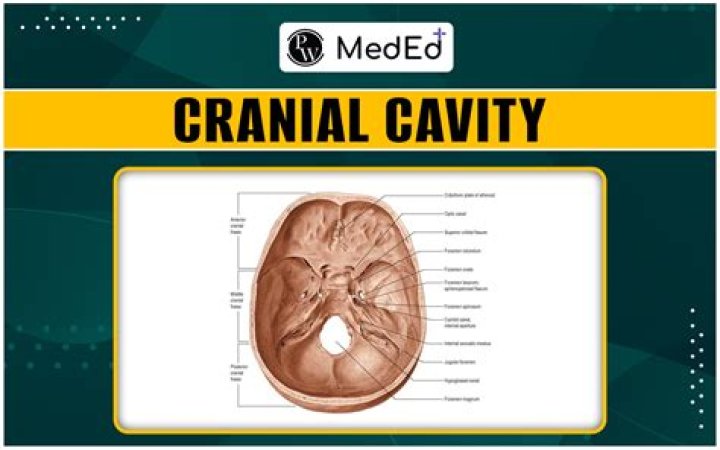
What structures form the boundaries of the cranial cavity

Ava Robinson
Published Apr 17, 2026
(b) The complex floor of the cranial cavity is formed by the frontal, ethmoid, sphenoid, temporal, and occipital bones. The lesser wing of the sphenoid bone separates the anterior and middle cranial fossae. The petrous ridge (petrous portion of temporal bone) separates the middle and posterior cranial fossae.
What are the boundaries of cranial cavity?
- Sphenoidal limbus (anterior margin of the chiasmatic groove)
- Posterior borders of the lesser wings of the sphenoid.
- Dorsum sellae of the sphenoid bone.
- Superior borders of the petrous part of the temporal bone.
- Groove for transverse sinus of the occipital bone.
Which bone forms part of the floor of the cranial cavity?
The ethmoid bone forms the central part of the floor, which is the deepest area of the anterior cranial fossa. In the center of this region is the cribriform plate, through which the olfactory tracts pass. The fovea ethmoidalis, or the roof of the ethmoid cavity, continues laterally from the cribriform plate.
What forms the wall of the cranial cavity?
The ethmoid bone forms the medial wall of the orbit, the roof of the nasal cavity, and due to its central location it articulates with numerous bones of the viscerocranium. Inside the neurocranium it articulates with the frontal and sphenoid bones.What bones form much of the walls and top of the cranial cavity?
The ethmoid bone is a single, midline bone that forms the roof and lateral walls of the upper nasal cavity, the upper portion of the nasal septum, and contributes to the medial wall of the orbit (Figure 6.23). On the interior of the skull, the ethmoid also forms a portion of the floor of the anterior cranial cavity.
What structures occupy anterior cranial fossa?
- frontal lobe of the cerebral cortex,
- olfactory bulb,
- olfactory tract,
- orbital gyri.
Which structures occupy the posterior cranial fossa?
The posterior cranial fossa is part of the cranial cavity, located between the foramen magnum and tentorium cerebelli. It contains the brainstem and cerebellum. This is the most inferior of the fossae. It houses the cerebellum, medulla and pons.
What structures are in the middle cranial fossa?
- superior orbital fissure.
- foramen rotundum.
- foramen ovale.
- foramen spinosum.
- foramen lacerum.
What are the boundaries of the middle cranial fossa?
- Sphenoidal limbus (anterior margin of the chiasmatic groove)
- Posterior borders of the lesser wings of the sphenoid.
- Dorsum sellae of the sphenoid bone.
- Superior borders of the petrous part of the temporal bone.
The coronal suture is a dense and fibrous association of connection tissue located in between the frontal and parietal bones of the skull. At birth, the sutures decrease in size (molding) and allow the skull to become smaller. In children, the suture enables the skull to expand with the rapidly growing brain.
Article first time published onWhich suture forms the boundary between the occipital bone and the parietal bones?
Lambdoid suture: the suture between the two parietal bones and the occipital bone.
Which of the following bones forms part of the lateral wall of the nasal cavity?
The lateral wall of the nasal cavity is formed partly by the maxilla, partly by the ethmoid bone, and partly by the perpendicular part of the palatine bone. Further back, where the nasal cavity becomes the nasopharynx, the lateral wall is formed by the medial pterygoid plate.
Which area of the brain is housed within the posterior cranial fossa?
Posterior Fossa This is a cavity in the back part of the skull which contains the cerebellum, brainstem and cranial nerves 5-12.
Which bone forms the greater portion of the sides and roof of the cranial cavity?
The two parietal bones form the greater portion of the sides and roof of the cranial cavity. The two temporal bones make up the lateral aspects of the cranium and part of the cranial floor. The zygomatic process of the temporal bone and the temporal process of the zygomatic bone form the zygomatic arch.
Which structures support and house the upper teeth in this bone?
The maxillary bone forms the upper jaw and supports the upper teeth. Each maxilla also forms the lateral floor of each orbit and the majority of the hard palate.
What structure is between the orbits of the eyes and superior to the bridge of the nose?
The lateral portions of the ethmoid bone are located between the orbit and upper nasal cavity, and thus form the lateral nasal cavity wall and a portion of the medial orbit wall.
What drains from posterior cranial fossa?
The straight sinus, receiving supra- and infratentorial Vein of Galen tributaries, the torcula, and both transverse-sigmoid (lateral) sinuses, represent the main drainage of the posterior fossa.
Which of the following connects the cranial cavity with the orbit?
Orbits are connected with the middle cranial fossa via the optic canal, the superior orbital fissure, the M-type orbitomeningeal foramen, the metoptic canal, an accessory anterior opening of the foramen rotundum, and Warwick’s canal.
Which structure occupies the posterior cranial fossa inferior to the cerebrum?
The cerebellum occupies the posterior fossa, dorsal to the pons and medulla. It is involved primarily in modulating motor control to enable precisely coordinated body movements. Similar to the cerebrum, which has gyri and sulci, the cerebellum has finer folia and fissures that increase the surface area.
What structures occupy the anterior cranial fossa middle cranial fossa?
The anterior, middle, and posterior cranial fossae house the anterior frontal lobe, temporal lobe, and cerebellum and brain stem, respectively.
What are Foramina of anterior cranial fossa?
Foramina/fissures of the anterior cranial fossa It lies in the frontal bone, just anterior to the ethmoid bone. It allows the passage of an emissary vein that comes from the nasal cavity and drains into the superior sagittal sinus, part of the venous drainage system associated with the brain.
What forms the floor of middle cranial fossa?
The dorsal boundary of the bony floor of the middle cranial fossa is formed by the anterior surface of the petrous part of the temporal bone. The floor of the middle fossa carries a number of portals for the passage of cranial nerves and vessels.
What Innervates the middle cranial fossa?
Previous neuroanatomical studies showed that the MMA in the dura mater along the floor of the middle cranial fossa is innervated by the NS originating from the mandibular and maxillary trigeminal divisions (Lv et al., 2014; Schueler et al., 2014).
What is the difference between fossa and cavity?
As nouns the difference between cavity and fossa is that cavity is a hole or hollow depression while fossa is (anatomy) a pit, groove, cavity, or depression, of greater or less depth or fossa can be a carnivorous mammal endemic to madagascar,.
What are the boundaries of the temporal fossa?
- Superiorly and posteriorly – superior temporal line.
- Inferiorly – zygomatic arch.
- Anteriorly – frontal process of the zygoma and zygomatic process of the frontal bone.
What separates anterior and middle cranial fossa?
The lesser wing of the sphenoid bone separates the anterior and middle cranial fossae. The petrous ridge (petrous portion of temporal bone) separates the middle and posterior cranial fossae.
What structure passes through the foramen magnum?
…a large oval opening, the foramen magnum, through which the medulla oblongata passes, linking the spinal cord and brain.
What type of joint is coronal suture?
The coronal suture is a dense, fibrous connective tissue joint that separates the two parietal bones from the frontal bone of the skull.
What flat bones form the coronal suture?
In the front, the parietal bones form the coronal suture with the frontal bone, and in the rear, the lambdoid suture is formed by the occipital bone. Finally, the squamosal suture separates the parietal and temporal bones.
What suture connects occipital and temporal bones?
The lambdoid suture is a line of dense, fibrous tissue that connects the occipital bone with the parietal bones. It is continuous with the occipitomastoid suture, which connects the occipital bone with the temporal bones.
Which suture forms the boundary between the temporal bone and the parietal bone of that side quizlet?
It is the largest fontanelle and lies at the intersection of the frontal, sagittal, and coronal sutures in the anterior portion of the skull. Where is the squamous suture located? On each side of the skull. Forms the boundary between the temporal bone and the parietal bone of that side.