What is the action of Pterygoids

Rachel Ross
Published Apr 24, 2026
The primary function of the lateral pterygoid
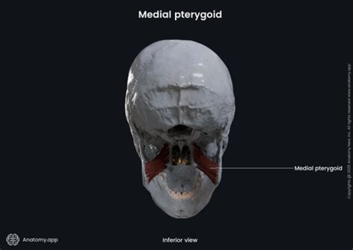
What is the main action of medial pterygoid?
The medial pterygoid muscle attaches to the angle of the mandible and to the lateral pterygoid plate to form a sling with the masseter muscle that suspends the mandible (Figure 6-19). The primary action is to elevate the mandible and laterally deviate it to the opposite side.
What muscle moves jaw forward?
Medial Pterygoid: This versatile muscle does triple duty. Two medial pterygoid muscles work on each side of your jaw: If you contract both muscles at once, your jaw moves forward.
What is the important action of the lateral pterygoid muscle?
Being a masticatory muscle, the lateral pterygoid aids in chewing and biting actions by controlling the movements of the mandible. The sphenoid attachment of the muscle is always fixed, meaning that the direction of pull is oriented towards it.What muscle is responsible for closing the mouth?
The masseter muscle is one of the four muscles responsible for the action of mastication (chewing). When the masseter contracts it causes powerful elevation of the mandible causing the mouth to close.
What is medial pterygoid plate?
The medial pterygoid plate (or medial pterygoid lamina) of the sphenoid bone is a horse-shoe shaped process that arises from its underside. … The anterior margin of the plate articulates with the posterior border of the vertical part of the palatine bone. In many animals it is a separate bone called the pterygoid bone.
Where does medial pterygoid attaches to the mandible?
OriginSuperficial part: Tuberosity of maxilla, Pyramidal process of palatine bone; Deep part: Medial surface of lateral pterygoid plate of sphenoid boneInsertionMedial surface of ramus and angle of mandible
What drains into pterygoid plexus?
In addition, the inferior ophthalmic vein and deep facial vein also drain into the pterygoid plexus. The plexus itself drains via the short maxillary vein before it forms the retromandibular vein. Emissary veins also anastomose between the plexus and the cavernous sinus, via the foramina ovale and lacerum.Why is the lateral pterygoid called the peripheral heart?
These communications are important for spread of infections and for collateral circulation. The pterygoid muscles and other muscles of mastication pump the blood from this plexus and are considered a “peripheral heart”. Chewing or yawning increases venous return.
What muscles do lateral deviation of the mandible?MusclesActionsLateral pterygoidProtracts mandible, depresses chin, lateral deviation of mandibleMedial pterygoidWorks with masseter to elevate mandible, aids in protrusion,Digastric Stylohyoid Mylohyoid GeniohyoidDepresses the mandible against resistance when infrahyoid muscles stabilize or depress hyoid bone
Article first time published onWhat are Pterygoids?
The pterygoid muscles are two of the four muscles of mastication, located in the infratemporal fossa of the skull. These muscles are: lateral pterygoid and medial pterygoid. The primary function of the pterygoid muscles is to produce movements of the mandible at the temporomandibular joint.
Why is the masseter so strong?
Strength in general usually refers to the capacity for either resistance or exertion. In the case of the masseter as the strongest muscle in the body it is because it can generate the largest measurable force of any single muscle. It is so strong for two main reasons. It is made up of densely packed muscle fibers.
Which muscle enables you to pucker your lips for a kiss?
Orbicularis oris: This muscle encircles your mouth and enables your lips to pucker for a kiss.
Is tongue a muscle?
Well, that’s only partly true: The tongue is really made up of many groups of muscles. These muscles run in different directions to carry out all the tongue’s jobs. The front part of the tongue is very flexible and can move around a lot, working with the teeth to create different types of words.
Is tongue the strongest muscle?
Many of us grew up believing the assertion that the tongue is the strongest muscle in the body. But is it really? The short answer is no.
What muscle is the strongest?
The strongest muscle based on its weight is the masseter. With all muscles of the jaw working together it can close the teeth with a force as great as 55 pounds (25 kilograms) on the incisors or 200 pounds (90.7 kilograms) on the molars.
What nerve innervates the medial pterygoid?
[2] The nerve to medial pterygoid is given off from the medial surface of the mandibular nerve. It travels through the otic ganglion (without synapsing) and gives motor innervation to the medial pterygoid muscle.
What type of joint is the temporomandibular joint?
The temporomandibular joint (TMJ), also known as the mandibular joint, is an ellipsoid variety of the right and left synovial joints forming a bicondylar articulation.
Which arteries supply blood to the medial and lateral pterygoid muscles?
Blood Supply and Lymphatics The medial pterygoid muscle receives vascular supply from the maxillary artery through its pterygoid branches and by the facial artery through its muscular branches. The pterygoid branches of the maxillary artery are 2 or 3 in number.
How do you examine medial pterygoid?
To palpate from outside the mouth, the head is tilted slightly to access the muscle. Palpation with one finger locates trigger points on the inner surface of the mandible by pressing upward at its angle. Palpation of the mid-belly is performed inside the mouth with the pad of the palpating index finger.
How does the peripheral heart work?
Together, the calf’s muscles and deep vein system form a complex array of valves and pumps, often referred to as the “peripheral heart,” that functions to push blood upward from the feet against gravity. The calf-muscle pump is analogous to the common hand-pump bulb of a sphygmomanometer filling a blood pressure cuff.
Which muscle is called peripheral heart and why?
Also, in upright posture, the soleus is responsible for pumping venous blood back into the heart from the periphery, and is often called the skeletal-muscle pump, peripheral heart or the sural (tricipital) pump. Soleus muscles have a higher proportion of slow muscle fibers than many other muscles.
What does the gastrocnemius and soleus do?
Function. Along with the soleus muscle, the gastrocnemius forms half of the calf muscle. Its function is plantar flexing the foot at the ankle joint and flexing the leg at the knee joint.
Why is pterygoid venous plexus important?
The function of the pterygoid venous plexus is to collect the blood from the palate, nasal cavity, paranasal sinuses, nasopharynx, and auditory tube. The veins of pterygoid plexus converge on its anterior end to form the maxillary vein, which conveys the blood from the plexus into the retromandibular vein.
Does pterygoid plexus drain into cavernous sinus?
The superior ophthalmic vein drains directly into the cavernous sinus through the superior orbital fissure. … The pterygoid plexus communicates with the cavernous sinus through the foramina ovale, spinosum, and rotundum.
What is a pterygoid plexus of veins?
The pterygoid venous plexus is an extensive valveless plexus of veins that parallels the medial two thirds of the maxillary artery on the lateral aspect of the medial pterygoid muscle, within the infratemporal fossa.
How does the mandible move?
Mandibular movement around the horizontal axis is an opening and closing motion. … When the condyles are in their most superior position in the articular fossae and the mouth is purely rotated open, the axis around which movement occurs is called the terminal hinge axis.
Which muscle is responsible when the mandible moves to the right?
Moving the mandible to one side (laterotrusion) involves contraction of the lateral pterygoid muscles on the opposite (contralateral) side from the excursion, assisted by the posterior temporalis muscle in the same side (ipsilateral).
What muscles elevate and retract the mandible?
The function of the masseter muscle is to elevate the mandible and approximate the teeth—additionally, the intermediate and deep muscle fibers of the masseter function to retract the mandible.
What muscle lowers the mandible?
Muscles that depress the mandible and thus open the jaw include the anterior digastric, mylohyoid, and inferior head of the lateral pterygoid. Jaw-closer muscles consist of the masseter, temporalis, medial pterygoid, and superior head of the lateral pterygoid.
What is the antagonist of the masseter?
The temporalis is the antagonist of the masseter. The temporalis is the insertion of the masseter. The temporalis is the origin of the masseter. Correct answer: … Since the temporalis assists in raising the jaw with the masseter, we say that the temporalis is the synergist of the masseter.