What is retinal pigment epithelium

Nathan Sanders
Published Apr 22, 2026
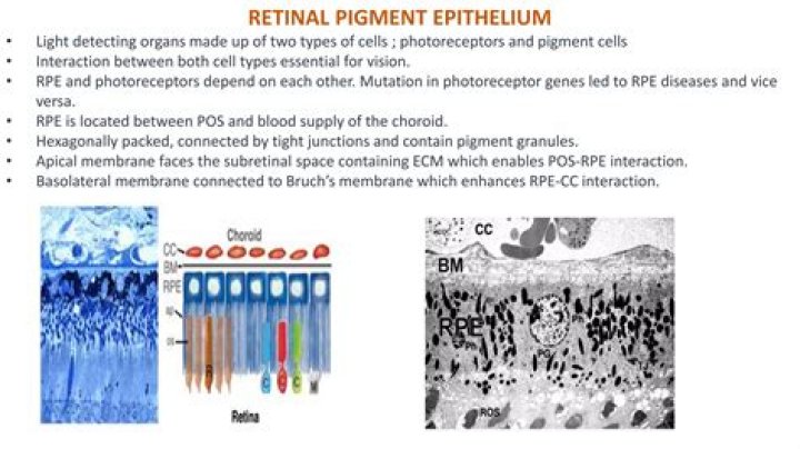
Retinal pigment epithelium (RPE) is formed from a single layer of regular polygonal cells arranged at the outermost layer of the retina. The outer side of the RPE is connected to Bruch’s membrane and the choroid, while the inner side is connected to the outer segment of photoreceptor cells.
What is the function of retinal pigment epithelium?
The retinal pigment epithelium (RPE) is a single layer of post-mitotic cells, which functions both as a selective barrier to and a vegetative regulator of the overlying photoreceptor layer, thereby playing a key role in its maintenance.
What is retinal pigment epithelial detachment?
Retinal pigment epithelial detachments (PEDs) are structural splitting within the inner aspect of Bruch’s membrane separating the retinal pigment epithelium (RPE) from the remaining Bruch’s membrane.
Where is retinal pigment epithelium?
The pigmented layer of retina or retinal pigment epithelium (RPE) is the pigmented cell layer just outside the neurosensory retina that nourishes retinal visual cells, and is firmly attached to the underlying choroid and overlying retinal visual cells.What is retinal pigment epithelial dystrophy?
Retinal pigment epithelial dystrophy (RPED) describes a disease of the retinal pigment epithelium, marked by the pathological accumulation of lipofuscin and associated with more widespread secondary retinal degeneration. This condition has also been referred to as “central progressive retinal atrophy” (CPRA).
What is the function of the retinal pigment epithelium quizlet?
∴ Epithelial cells are heavily pigmented to prevent light from entering the eye, except through the pupil and reduce scatter.
What causes retinal pigmentation?
What causes RP? RP is an inherited disorder that results from harmful changes in any one of more than 50 genes. These genes carry the instructions for making proteins that are needed in cells within the retina, called photoreceptors.
Does the retinal pigment epithelium release glutamate?
Thus, glutamate released by RPE cells may influence synaptic transmission at the main signaling pathway of the retina. Although glutamate release from photoreceptors to inner retinal neurons has been extensively documented, the role of glutamate in communicating the RPE with the neural retina is largely unexplored.What is retinal pigment epithelial mottling of macula?
Definition. Mottling (spots or blotches with different shades) of the retinal pigment epithelium, i.e., localized or generalized fundal pigment granularity associated with processes at the level of the retinal pigment epithelium. [
What do pigment cells do?One function of pigment cells may be to eradicate oxygen radicals that are responsible in part for inducing malignancies and are also involved in the aging process.
Article first time published onWhat is the treatment for pigment epithelial detachment?
Pigment epithelial detachment resolution was demonstrated in a significant proportion of eyes treated with ranibizumab or aflibercept therapy. There was additional resolution with a higher dose of anti-VEGF (ranibizumab 2.0 mg) versus a lower dose (ranibizumab 0.5 mg).
What causes a pigment epithelial detachment?
Pigment epithelial detachment is a condition that happens when specific layers of cells behind your eye come apart, or get detached. This kind of detachment happens when you have extra fluid or other material under a layer of cells in the back of your eye, called the retinal pigment epithelium (RPE).
Is PED serious?
In AMD, PEDs are important markers of disease severity, risk for progression, and vision loss. PEDs are seen in up to 62% of eyes with advanced AMD,4 and approximately 50% of patients with newly diagnosed PEDs will experience significant visual loss (>3 lines) one year from diagnosis.
What causes Metamorphopsia?
They’re caused by a defect in the surface lining of the retina. This defect can be caused by age, retinal tears, and diseases like diabetes, which affect vascular regions in the eye. ERMs begin by cells growing on the smooth retinal membrane.
Is there a cure for cone-rod dystrophy?
Currently, there is no treatment to stop a person with cone-rod dystrophy (CRD) from losing their vision. However, there may be treatment options that can help slow down the degenerative process, such as light avoidance and the use of low-vision aids.
What causes pattern dystrophy?
Pattern dystrophies are caused by mutations (or mistakes) in one of several genes, but they are all inherited in an autosomal dominant fashion. That means that someone with a pattern dystrophy has a 50 per cent chance of passing it on to their child, whether they are male or female.
Does retinitis pigmentosa always lead to blindness?
Retinitis Pigmentosa Symptoms Symptoms vary, depending on the type of retinal cell that is affected. Both eyes often experience similar vision loss. It should be noted that RP is a slowly progressive disease over many years and that most patients never become completely blind.
How long is the average lifespan of a person with retinitis pigmentosa?
Patients with this amplitude are expected to retain some useful vision for their entire lives assuming an average life expectancy of 80 years. With vitamin A treatment the critical voltage appears to be 2 μV or greater at age 40.
At what age does retinitis pigmentosa occur?
RP is typically diagnosed in young adulthood, but the age of onset may range from early childhood to the mid 30s to 50s. Photoreceptor degeneration has been detected as early as age of six years even in patients who remain asymptomatic until young adulthood.
Which of the following is the function of the pigmented layer of the eye quizlet?
The outer layer that is composed of only a single layer of pigment cells (melanocytes). Absorbs light and prevents it from scattering in the eye. Pigment cells act as phagocytes for cleaning up cell debris and also store vitamin A needed for photorecptor renewal.
What causes macular mottling?
Your eye doctor will put drops in your eyes to dilate them and use a special instrument to examine the back of your eye. He or she will look for a mottled appearance that’s caused by drusen – yellow deposits that form under the retina. People with macular degeneration often have many drusen.
What does pigment changes in the eye mean?
While most people develop some very small drusen as a normal part of aging, the presence of medium-to-large drusen may indicate that you have macular degeneration. Another sign of macular degeneration is the appearance of pigment changes in the retina.
Is retinal vascular or avascular?
The retina of lower vertebrates is avascular, but is usually provided with two external blood supplies, the choriocapillaris on the outer surface and a vitreal blood supply on the inner surface.
How is this pigment important to eye function?
Melanin granules in the retinal pigment epithelium (RPE) have many important functions which are not yet completely understood. Melanin in the RPE protects the cell from damage caused by oxidative stress. … Thus, melanin protects against light toxicity and against cytotoxic effects caused by ocular inflammation.
Where is the epithelium?
The epithelium is a type of body tissue that forms the covering on all internal and external surfaces of your body, lines body cavities and hollow organs and is the major tissue in glands.
What are pigment cells called?
Chromatophores are cells that produce color, of which many types are pigment-containing cells, or groups of cells, found in a wide range of animals including amphibians, fish, reptiles, crustaceans and cephalopods. Mammals and birds, in contrast, have a class of cells called melanocytes for coloration.
Where is melanin produced?
Melanin is formed primarily in the melanocyte, located in the inner layers of the skin where melanin and carotene blend to produce the skin color as well as the color in the eyes and hair. Red hair is produced by pheomelanin in spherical melanosomes (melanin granules).
How do you get pigment cells?
There aren’t any studies that prove vitamin C increases melanin production. However, anecdotal evidence suggests vitamin C might increase melanin levels. Eating vitamin C–rich foods like citrus, berries, and leafy green vegetables may optimize melanin production. Taking a vitamin C supplement may help as well.
Is central serous retinopathy curable?
Central Serous Retinopathy most commonly presents as a one-time event in just one eye and resolves spontaneously without treatment within three to four months. CSR can occasionally occur in both eyes. In some people, it can recur unpredictably months or years after the initial episode has resolved.
What is CSR eye disease?
Central serous retinopathy (CSR) or central serous chorioretinopathy (CSCR) affects the central area of your retina known as the macula. CSR can cause your vision to be blurred and distorted due to fluid collecting underneath your macula.
Can you go blind from a detached retina?
If the retinal detachment isn’t treated right away, more of the retina can detach — which increases the risk of permanent vision loss or blindness.