What goes through the foramen of the skull

Zoe Patterson
Published Apr 17, 2026
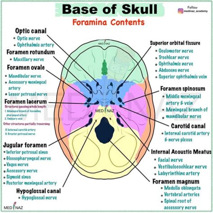
The human skull has numerous foramina through which cranial nerves, arteries, veins, and other structures pass. The skull bones that contain foramina include the frontal, ethmoid, sphenoid, maxilla, palatine, temporal, and occipital lobes.
What cranial nerves go through what foramen?
LocationNerveforamen ovaleTrigeminal V3 (mandibular)stylomastoid foramenFacial nerve (VII)internal auditory canalVestibulocochlear (VIII)jugular foramenGlossopharyngeal (IX) Vagus (X) Accessory (XI)
What are the foramen?
The foramen is the bony hollow archway created by pedicles of adjacent vertebrae, creating a passageway through which all spinal nerve roots run. As a spinal nerve branches from the spinal cord, it exits through this opening and travels to organs, muscles and sensory structures of the body.
What exits through the foramen ovale?
The following structures pass through foramen ovale: mandibular nerve, a branch of the trigeminal nerve. accessory meningeal artery. lesser petrosal nerve, a branch of the glossopharyngeal nerve.What passes through the foramen cecum?
The foramen cecum varies in size in different subjects, and is frequently impervious; when open, it transmits the emissary vein from the nose to the superior sagittal sinus.
Which of the following nerves exits the skull through the foramen ovale?
The mandibular nerve (V3) exits the cranium through the foramen ovale (Figures 2.2 and 2.3). On its extracranial course, it divides into three main branches: the buccal, mental, and auriculotemporal nerves.
What goes through jugular foramen?
The structures that traverse the jugular foramen are the sigmoid sinus and jugular bulb, the inferior petrosal sinus, meningeal branches of the ascending pharyngeal and occipital arteries, the glossopharyngeal, vagus, and accessory nerves with their ganglia, the tympanic branch of the glossopharyngeal nerve (Jacobson’s …
What goes through the cranial fossa?
The human skull has numerous openings (foramina), through which cranial nerves, arteries, veins, and other structures pass. These foramina vary in size and number, with age.What is the foramen ovale in the skull?
The foramen ovale is another opening located at the base of the greater wing of the sphenoid. It is positioned posterolateral to the foramen rotundum within the middle cranial fossa. It conducts the mandibular nerve (branch of the trigeminal nerve, CN V) and the accessory meningeal artery.
Where is the foramen located?Foramen magnumPart ofoccipital boneSystemskeletalIdentifiersLatinForamen magnum
Article first time published onWhat is the foramen magnum?
The foramen magnum is the largest foramen of the skull. It is located in the most inferior portion of the cranial fossa as a part of the occipital bone. … On the foramen magnum, there are two craniometric points: the basion, the median point of the front edge of the hole, and the opisthion, posterior correspondence.
Where are the sutures of the skull?
- Metopic suture. This extends from the top of the head down the middle of the forehead, toward the nose. …
- Coronal suture. This extends from ear to ear. …
- Sagittal suture. …
- Lambdoid suture.
What nerve passes through the foramen Rotundum?
The maxillary nerve (V2) passes through the foramen rotundum and into the infraorbital canal, where, at the pterygopalatine fossa, it branches into the pterygopalatine ganglion, with parasympathetic and sensory branches to the paranasal sinuses.
What nerves pass through foramen magnum?
Cranial nerves: cranial nerves IX and X also pass through the foramen magnum. The ninth cranial nerve is the glossopharyngeal nerve that begins at the medulla oblongata; the tenth cranial nerve is the vagus nerve – probably the most broadly functioning cranial nerve.
What goes through hypoglossal canal?
Cranial nerves These nerves originate in the motor nuclei of the medulla, passing through the hypoglossal canals of the occipital bone, to reach the tongue muscles. Each hypoglossal nerve exits the cranium and curves, reaching the skeletal tongue muscles. It provides voluntary motor control of tongue movements.
What passes through the carotid foramen?
The bony canal that extends rostrally from the caudal foramen lacerum is called the carotid canal; it transmits the internal carotid artery and postganglionic sympathetic plexus from the superior cervical ganglion (also called the carotid plexus, made up of the carotid nerves; Figure 1-11).
What is the Glossopharyngeal?
Introduction. The glossopharyngeal nerve is the 9th cranial nerve (CN IX). It is one of the four cranial nerves that has sensory, motor, and parasympathetic functions. It originates from the medulla oblongata and terminates in the pharynx.
What passes through the optic foramen quizlet?
The optic foramen, through which the CNII and Opthalmic artery branch of ICA go through, are on the lesser wing of the sphenoid and open up into the optic canal.
Where is the foramen ovale?
What is patent foramen ovale? A patent foramen ovale (PFO) is a small opening between the two upper chambers of the heart, the right and the left atrium. Normally, a thin membranous wall made up of two connecting flaps separates these chambers. No blood can flow between them.
What is foramen cecum?
The foramen cecum represents a primitive tract between the anterior cranial fossa and the nasal space. It is located along the anterior cranial fossa, anterior to the cribriform plate of the ethmoid bone and posterior to the frontal bone, within the frontoethmoidal suture.
Where is the cranial fossa?
A cranial fossa is formed by the floor of the cranial cavity. There are three distinct cranial fossae: Anterior cranial fossa (fossa cranii anterior), housing the projecting frontal lobes of the brain. Middle cranial fossa (fossa cranii media), separated from the posterior fossa by the clivus and the petrous crest.
What's the difference between foramen and foramina?
The intervertebral foramen, also called the neural foramen, is the opening between the vertebrae through which spinal nerve roots travel and exit to other parts of the body. The word “foramen” is the singular form, while “foramina” is the plural form.
What is the function of foramen?
Foramina inside the body of animals typically allow muscles, nerves, arteries, veins, or other structures to connect one part of the body with another.
What is the largest foramen in the skull?
The foramen magnum is the largest foramen of the skull. It is located in the most inferior portion of the cranial fossa as a part of the occipital bone.
What is a foramen in a bone?
Foramen – A hole through which nerves and blood vessels pass. Examples include supraorbital foramen, infraorbital foramen, and mental foramen on the cranium. Fossa – A shallow depression in the bone surface. … It is separated from the shaft of the bone by the neck.
What is spiral cord?
The spinal cord is a long bundle of nerves and cells that extends from the lower portion of the brain to the lower back. It carries signals between the brain and the rest of the body.
What level is foramen magnum?
The foramen magnum is found in the most inferior part of the posterior cranial fossa 3. It is traversed by vital structures including the medulla oblongata 1. In the midline, the anterior margin is the basion and the posterior margin is the opisthion.
What is Infraorbital foramen?
The infraorbital foramen, an opening into the floor of the eye socket, is the forward end of a canal through which passes the infraorbital branch of the maxillary nerve, the second division of the fifth cranial nerve.
What are the 5 sutures of the skull?
The main sutures of the skull are the coronal, sagittal, lambdoid and squamosal sutures. The metopic suture (or frontal suture) is variably present in adults.
What are the 6 sutures of the skull?
Six primary sutures of the cranial vault exist, including the paired coronal sutures (between the frontal and parietal bones), the paired lambdoid sutures (between the parietal and interparietal bones), the single sagittal suture (between the parietal bones), and the single human metopic or murine posterior frontal …
Where is maxillary?
The maxilla is the bone that forms your upper jaw. The right and left halves of the maxilla are irregularly shaped bones that fuse together in the middle of the skull, below the nose, in an area known as the intermaxillary suture.