What do tall T waves mean

Samuel Coleman
Published Apr 19, 2026
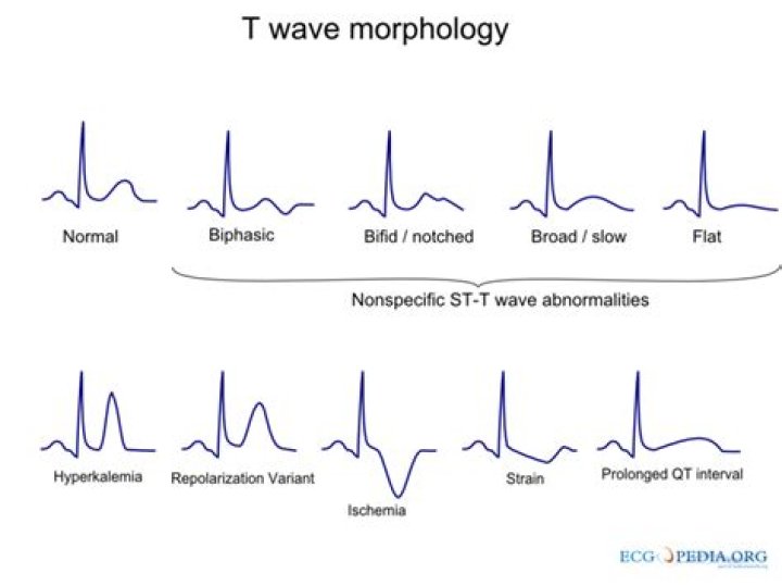
Tall T-waves (also called hyper-acute T waves) can be an early sign of ST-elevation myocardial infarction. The morphology of the T waves can begin to broaden and peak within 30 minutes of complete coronary artery occlusion, and thus may be the earliest sign of myocardial infarction on the EKG.
What does a high T wave indicate?
For example, tall, peaked T waves in a patient who missed three runs of dialysis are likely to represent hyperkalemia, while tall “hyperacute” T waves in a patient complaining of the acute onset of crushing, sub-sternal chest pain could represent the acute onset of transmural myocardial ischemia.
Is Tall T wave is normal?
In normal adults, T wave inversions are less commonly found, but can be normal from V1 to V3. The depth of the T wave also becomes progressively shallow from one to the next lead. The height of the T wave should not exceed 5 mm in limb leads and more than 10 mm in precordial leads.
When do you use tall T waves?
Narrow and tall peaked T wave (A) is an early sign of hyperkalemia. It is unusual for T waves to be taller than 5 mm in limb leads and taller than 10 mm in chest leads. Hyperkalemia should be suspect if these limits are exceeded in more than one lead.Can anxiety cause Tall T waves?
A study by Whang et al. (2014) showed that depressive and anxious symptoms were associated with abnormalities in T wave inversions.
Can hyperventilation cause an elevated T wave?
Hyperventilation is already known as a cause of transient T-wave inversion; however, it is often forgotten in modern clinical settings.
Should I worry about abnormal T wave?
So, my advice to you is not to worry. Inverted T-waves are not uncommon, and you don’t need to be overly anxious about them as long as you continue to feel well and have normal echocardiograms and stress tests.
Why is the T wave upright on an ECG?
Generally, the T wave exhibits a positive deflection. The reason for this is that the last cells to depolarize in the ventricles are the first to repolarize.Can stress cause T waves?
Abnormal ECG Findings Caused by Anxiety Whether it is due to short-term test nervousness or a chronic condition, anxiety may be associated with certain ECG abnormalities, including T-wave inversion.
What is ischemia of the heart?Myocardial ischemia occurs when blood flow to the heart muscle (myocardium) is obstructed by a partial or complete blockage of a coronary artery by a buildup of plaques (atherosclerosis). If the plaques rupture, you can have a heart attack (myocardial infarction).
Article first time published onIs a sinus rhythm good?
It means the electrical pulse from your sinus node is being properly transmitted throughout the heart muscle. In adults, normal sinus rhythm usually accompanies a heart rate of 60 to 100 bpm. However, it’s possible for sinus rhythm to be faster or slower than this and still be considered normal.
What causes an inverted T-wave?
T wave inversions in the right chest leads may be caused by right ventricular overload (e.g., acute or chronic pulmonary embolism) and in the left chest leads by left ventricular overload (Chapter 7). Diffusely inverted T waves are seen during the evolving phase of pericarditis or myocarditis.
Can high blood pressure cause an abnormal EKG?
High blood pressure Other aspects of heart disease may lead to an abnormal EKG. For example, people with high blood pressure are more likely to have an abnormal EKG reading.
What causes ST and T wave abnormality?
“Primary” ST-T Wave Abnormalities (ST-T wave changes that are independent of changes in ventricular activation and that may be the result of global or segmental pathologic processes that affect ventricular repolarization): Drug effects (e.g., digoxin, quinidine, etc) Electrolyte abnormalities (e.g., hypokalemia)
Can dehydration cause abnormal EKG?
For example, a person with dehydration may have imbalanced electrolytes that are causing an abnormal EKG. This person may require fluids, electrolyte-containing beverages, or medications to restore electrolytes. Sometimes, a doctor may not recommend any treatments for an abnormal EKG.
What are the symptoms of hyperventilation syndrome?
- Dizziness or lightheadedness.
- Shortness of breath.
- Belching, bloating, dry mouth.
- Weakness, confusion.
- Sleep disturbances.
- Numbness and tingling in your arms or around your mouth.
- Muscle spasms in hands and feet, chest pain and palpitations.
Can hyperventilation cause ST depression?
These findings suggest that early hyperventilation-induced ST segment depression is due to increased oxygen demand in patients with poor coronary reserve and may be prevented by beta-adrenergic blockers, which are useful for lowering oxygen consumption.
Can anxiety cause an inverted T-wave?
(HealthDay)—Depression and anxiety are independently, yet oppositely, associated with electrocardiographic (ECG) T-wave inversions, according to a study published in the Dec. 15 issue of The American Journal of Cardiology.
How long is a normal T wave?
Often the onset and end of the T Wave are difficult to determine with certainty. The DURATION of the T Wave is 0.10 to 0.25 seconds or greater. The AMPLITUDE of the T Wave is less than 5 mm. The SHAPE of the T Wave is sharply or bluntly rounded and slightly asymmetrical.
What does ischemia feel like?
What are symptoms of myocardial ischemia? The most common symptom of myocardial ischemia is angina (also called angina pectoris). Angina is chest pain that is also described as chest discomfort, heaviness, tightness, pressure, aching, burning, numbness, fullness, or squeezing. It can feel like indigestion or heartburn.
What are 3 tests that help recognize heart disease?
- Electrocardiogram (ECG or EKG). An ECG is a quick and painless test that records the electrical signals in your heart. …
- Holter monitoring. …
- Echocardiogram. …
- Stress test. …
- Cardiac catheterization. …
- Cardiac computerized tomography (CT) scan. …
- Cardiac magnetic resonance imaging (MRI).
Can Ischaemic heart disease cause sudden death?
It is well known that ischemic heart disease is the leading cause of sudden death, being responsible for more than 80% of cases.
Can your sinuses affect your heart?
The sinus node can increase your heart rate during times of high demand, such as during exercise. If you have sick sinus syndrome, your heart rate may be too slow or too fast to meet the needs of your body. About 1 in 600 people with heart problems have sick sinus syndrome.
Is sinus arrhythmia serious?
Keep in mind that for the majority of people, a sinus arrhythmia is neither dangerous nor problematic. Even if your doctor suspects you have this irregular heartbeat, he may not order the test to check for it. That’s because an EKG can be costly, and a sinus arrhythmia is considered a benign condition.
Should I be worried about sinus arrhythmia?
Respiratory sinus arrhythmia is not considered a major health concern. However, other arrhythmias can sometimes indicate heart disease. An older person with a severe arrhythmia may require a pacemaker.
What do inverted T waves look like?
In patients with implanted right ventricular pacemakers, inverted T waves are most often seen in leads I and aVL. The T waves are inverted in an asymmetric fashion with a gradual initial downslope and an abrupt return to the baseline.
Would an EKG show a blockage?
An ECG Can Recognize the Signs of Blocked Arteries. Unfortunately, the accuracy of diagnosing blocked arteries further from the heart when using an ECG decrease, so your cardiologist may recommend an ultrasound, which is a non-invasive test, like a carotid ultrasound, to check for blockages in the extremities or neck.
How do I know my heart is failing?
Heart failure signs and symptoms may include: Shortness of breath with activity or when lying down. Fatigue and weakness. Swelling in the legs, ankles and feet.
Can an ECG detect a blocked artery?
No, an electrocardiogram cannot detect blocked arteries. Blocked arteries are usually diagnosed with a nuclear stress test, cardiac pet scan, coronary CT angiogram or traditional coronary angiogram.