Is this blood accumulation above the dura mater or below the dura mater

Ava Robinson
Published Apr 23, 2026
Bleeding between the dura and bone is called an epidural hematoma
Is the blood accumulation above or below the dura mater?
A subdural hematoma is a type of brain bleed. Blood leaks out of a blood vessel into the space below the outermost membrane of the brain — the dura mater.
What is the medical term for above the dura?
Epidural means outside the dura. An accumulation of blood outside the dura is an epidural hematoma.
Where does blood accumulate in a subdural hematoma?
In a subdural hematoma, the blood seeps between the dura and the arachnoid layers. It collects inside the brain’s tough outer lining. This bleeding often comes from a blood vessel that breaks within the space around the brain. This most often happens because of a head injury.Which type of bleed is between the skull and the dura mater?
Also called an extradural hematoma, this type occurs when a blood vessel — usually an artery — ruptures between the outer surface of the dura mater and the skull. Blood then leaks between the dura mater and the skull to form a mass that presses on brain tissue. The most common cause of an epidural hematoma is trauma.
What is hematoma?
A hematoma is a bad bruise. It happens when an injury causes blood to collect and pool under the skin. The pooling blood gives the skin a spongy, rubbery, lumpy feel. A hematoma usually is not a cause for concern. It is not the same thing as a blood clot in a vein, and it does not cause blood clots.
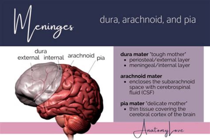
What is the dura?
(DER-uh MAY-ter) The tough outer layer of tissue that covers and protects the brain and spinal cord and is closest to the skull. The dura mater is one of the three layers that form the meninges.
Does subdural hematoma cause increased ICP?
As a subdural hematoma expands in the subdural space, it raises the ICP and deforms the brain. The rise in ICP is initially compensated by efflux of cerebrospinal fluid (CSF) toward the spinal axis and compression of the venous system, expediting venous drainage through the jugular veins.Is subdural hematoma venous or arterial?
Subdural hematomas are usually of venous origin and progress slowly, as opposed to epidural hematomas that are of arterial origin and may reach maximum size within minutes.
Which vessel is involved in subdural hematoma?Acute subdural hematoma is usually caused by external trauma that creates tension in the wall of a bridging vein as it passes between the arachnoid and dural layers of the brain’s lining—i.e., the subdural space. The circumferential arrangement of collagen surrounding the vein makes it susceptible to such tearing.
Article first time published onWhy is it called the dura mater?
The name dura mater derives from the Latin for tough mother (or hard mother), a loan translation of Arabic أم الدماغ الصفيقة (umm al-dimāgh al-ṣafīqah), literally ‘thick mother of the brain’, matrix of the brain, and is also referred to by the term “pachymeninx” (plural “pachymeninges”).
What does the dura mater do?
The dura mater is a sac that envelops the arachnoid and has been modified to serve several functions. The dura mater surrounds and supports the large venous channels (dural sinuses) carrying blood from the brain toward the heart. The dura mater is partitioned into several septa, which support the brain.
Why is it called arachnoid mater?
The arachnoid mater, named for its spiderweb-like appearance, is a thin, transparent membrane surrounding the spinal cord like a loosely fitting sac. Continuous with the cerebral arachnoid above, it passes through the foramen magnum and descends caudally to the S2 vertebral level.
What are the 4 types of brain bleed?
Intracranial hemorrhage encompasses four broad types of hemorrhage: epidural hemorrhage, subdural hemorrhage, subarachnoid hemorrhage, and intraparenchymal hemorrhage. [1][2][3] Each type of hemorrhage is different concerning etiology, findings, prognosis, and outcome.
What is a bleed on the brain called?
Bleeding in the brain (also called a brain hemorrhage or brain bleed) can happen because of an accident, brain tumor, stroke, or high blood pressure caused by congenital or other health conditions.
What is an epidural bleed?
An epidural hematoma (EDH) is bleeding between the inside of the skull and the outer covering of the brain (called the dura).
Is dura mater avascular?
Epidural Space and Dura Mater While it was once thought that the dura was avascular, it is actually highly vascular in nature, as the major vessels that supply it run in the epidural space deep to the skull.
Does dura mater have blood vessels?
Blood Supply and Lymphatics The dura mater receives vascular supply from the following branches: Internal carotid artery. Maxillary artery. Ascending pharyngeal artery.
What is the dura mater made up of?
The dura mater is made up of fibroblasts and large amounts of extracellular collagen. The dura mater is composed of two layers: the periosteal/endosteal layer and the meningeal layer. The dural venous sinuses are between these two layers.
Is a hematoma the same as a blood clot?
Blood clots typically occur inside larger blood vessels, such as an artery or a vein. Doctors refer to a blood clot as a thrombus. Damage to blood vessels can cause large amounts of blood to leak into the surrounding tissue, forming so-called hematoma. This collection of blood can then become sticky and harden.
What is a scalp hematoma?
Scalp hematoma: A scalp hematoma typically appears as a bump on the head. The damage is to the external skin and muscle, so it will not affect the brain. Septal hematoma: Usually the result of a broken nose, a septal hematoma may cause nasal problems if a person does not receive treatment.
What is the difference between a bruise and a hematoma?
A bruise, also known as a contusion, typically appears on the skin after trauma such as a blow to the body. It occurs when the small veins and capillaries under the skin break. A hematoma is a collection (or pooling) of blood outside the blood vessel.
Is subarachnoid hemorrhage arterial or venous?
A subarachnoid hemorrhage is bleeding from a damaged artery at the surface of the brain. This bleeding often causes a sudden, severe headache. It is a medical emergency. Subarachnoid hemorrhage is a type of stroke.
What causes increased ICP?
Increased ICP can result from bleeding in the brain, a tumor, stroke, aneurysm, high blood pressure, or brain infection. Treatment focuses on lowering increased intracranial pressure around the brain. Increased ICP has serious complications, including long-term (permanent) brain damage and death.
What causes increased CSF?
Common causes include: Aneurysm rupture and subarachnoid hemorrhage. Brain tumor. Encephalitis irritation and swelling, or inflammation, of the brain)
What causes increased CSF production?
The increased CSF production is the result of an increased activity of Na+-K+ ATPase at the choroid plexus level, which establishes a sodium gradient across the choroid epithelial cells, as well as of an elevated CBF (66).
What is the pathophysiology of a subdural hematoma?
An acute subdural hematoma (SDH) is a clot of blood that develops between the surface of the brain and the dura mater, the brain’s tough outer covering, usually due to stretching and tearing of veins on the brain’s surface. These veins rupture when a head injury suddenly jolts or shakes the brain.
Where are the bridging veins?
Bridging veins are veins in the subarachnoid space that puncture the dura mater and empty into the dural venous sinuses. A rupture of a bridging vein causes a subdural hematoma.
Where is the dura in the brain?
Dura mater is on the outermost end of the meninges, situated directly beneath the skull and the bones of the vertebral column. The dura mater is made up of two layers of connective tissue: the periosteal layer and the meningeal layer. The periosteal layer lines the inner surface of the cranium’s bones.
What area of the brain controls blood pressure?
The brain stem sits beneath your cerebrum in front of your cerebellum. It connects the brain to the spinal cord and controls automatic functions such as breathing, digestion, heart rate and blood pressure.
What are the reflections of dura mater Invaginated between parts of the brain and where are they situated?
The dura mater is a two-layered membrane attached to the inner surface of the skull [1,2]. … The dural reflections of the 2 meningeal layers fold and invaginate into the cranial cavity by forming the falx cerebri and the tentorium cerebelli.