Is basilar artery part of circle of Willis

Andrew Mitchell
Published Apr 19, 2026
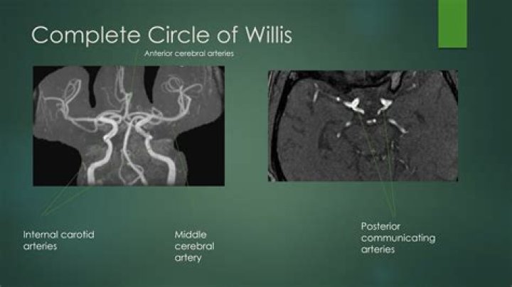
The anterior communicating, anterior cerebral, internal carotid, posterior communicating, posterior cerebral, and basilar arteries are all part of the circle of Willis (see Fig.
How many arteries are in circle of Willis?
The circle of Willis is a group of blood vessels in the brain that connect with each other, forming a continuous structure that resembles a circle. These nine arteries supply blood to a large portion of the brain. Most of the time, blood can flow through the vessels of the circle of Willis without any interruption.
Where is basilar artery located?
The basilar artery is part of the blood supply system for the brain and central nervous system. It is formed where the two vertebral arteries join at the base of the skull. The basilar artery carries oxygenated blood to the cerebellum, brainstem, and occipital lobes.
Is basilar artery in circle of Willis?
Origin of arteries The anterior cerebral artery forms the anterolateral portion of the circle of Willis, while the middle cerebral artery does not contribute to the circle. The right and left posterior cerebral arteries arise from the basilar artery, which is formed by the left and right vertebral arteries.Which arteries feed the circle of Willis?
Two arteries, called the carotid arteries, supply blood to the brain. They run along either side of the neck and lead directly to the circle of Willis. Each carotid artery branches into an internal and external carotid artery. The internal carotid artery then branches into the cerebral arteries.
Which arteries form the circle of Willis quizlet?
- Posterior Cerebral Artery. – From Vertebral Artery.
- Posterior Communicating Artery. – Connects Posterior Cerebral to Internal Carotid.
- Internal Carotid Artery.
- Anterior Cerebral Artery. – from Internal Carotid.
- Anterior communication artery.
What constitutes the circle of Willis?
The Circle of Willis is the joining area of several arteries at the bottom (inferior) side of the brain. At the Circle of Willis, the internal carotid arteries branch into smaller arteries that supply oxygenated blood to over 80% of the cerebrum.
What is a fenestrated basilar artery?
Fenestration of a cerebral artery is a type of rare congenital vascular dysplasia. It is characterised by the division of an artery, with two distinct endothelium-lined channels within the lumen of a single artery. 1 Among cerebral arteries, basilar artery fenestration is the most common form of fenestration.Who discovered circle of Willis?
Thomas Willis (1621-1675) (Figure 7) is best known for his description and figuration of the circle of Willis.
What is Hyperdense basilar artery?The hyperdense basilar artery sign (HBAS) is an indicator of vessel occlusion on non contrast-enhanced computer tomography (NECT) in acute stroke patients. Since basilar artery occlusion (BAO) is associated with a high mortality and morbidity, its early detection is of great clinical value.
Article first time published onWhat 2 arteries give rise to the basilar artery?
The basilar artery is a midline structure formed from the confluence of the vertebral arteries. Terminally, the basilar artery branches to establish the right and left posterior cerebral arteries.
Why is the circle of Willis important?
The circle of Willis acts to provide collateral blood flow between the anterior and posterior circulations of the brain, protecting against ischemia in the event of vessel disease or damage in one or more areas.
What is an incomplete circle of Willis?
The circle of Willis (CoW) is considered an important collateral network to maintain blood flow when some of the supply is diminished. Previous studies showed that CoW is incomplete in approximately 50% to 90% of adults and the number of missing segments correlates with the intolerance to cross-clamping.
Which artery is not part of the cerebral arterial circle?
The basilar artery and middle cerebral arteries, though they supply the brain, are not considered part of the circle.
What is the ophthalmic artery?
The ophthalmic artery is the first branch off of the internal carotid artery and supplies the optic nerve and retina. From: Essential Emergency Medicine, 2007.
What part of the circle of Willis is the most common site of aneurysm?
Most cerebral aneurysms are found at predictable locations around the circle of Willis; the three most common are the junction of the anterior communicating artery with the anterior cerebral artery (30% to 35%), the posterior communicating artery at the junction with the internal carotid artery (30% to 35%), and the …
Which arteries communicate with the basilar artery to form the circle of Willis quizlet?
Both pairs originate in turn from subclavian arteries (branches of the brachio-cephalic trunk of the aorta). As two vertebral arteries climb via inter-vertebral foramena toward the brain, they unite on the frontal surface of the pons and form BASILAR ARTERY.
Which artery does not form the circle of Willis quizlet?
Terms in this set (10) The basilar artery and middle cerebral arteries, supplying the brain, are not considered part of the circle.
What is the circle of Willis and why is it important quizlet?
The circle of willis is an important means of collateral circulation in the event of gradual obstruction of one of the major arteries forming the circle.
What's the main artery called?
The largest artery is the aorta, the main high-pressure pipeline connected to the heart’s left ventricle. The aorta branches into a network of smaller arteries that extend throughout the body. The arteries’ smaller branches are called arterioles and capillaries.
What is the communicating artery?
In human anatomy, the anterior communicating artery is a blood vessel of the brain that connects the left and right anterior cerebral arteries.
How can I memorize my arteries?
A useful mnemonic to remember the branches of the internal iliac artery is: I Love Going Places In My Very Own Underwear! Mnemonic I: iliolumbar artery L: lateral sacral artery G: gluteal (superior and inferior) arteries P: (internal) pudendal artery I: inferior vesical (vaginal in female…
Which artery supplies blood to the lower lip?
We found that the blood supply of the lower lip was derived from the facial artery and three dominant labial arteries: the inferior labial artery, the horizontal labiomental artery, and the vertical labiomental artery.
Which of the following arteries does not feed directly or indirectly into the cerebral arterial circle circle of Willis )?
Anatomy 2 Cardiovascular system: Blood Vessels and Circulation Flashcards | Quizlet.
What is subclavian artery?
The subclavian arteries lie just below the clavicles, providing blood supply to the bilateral upper extremities with contributions to the head and neck. The right subclavian artery derives from the brachiocephalic trunk, while the left subclavian artery originates directly from the aortic arch.
What is a fenestration procedure?
Fenestration closure after a Fontan operation is a procedure to close the hole between your child’s heart and the tunnel that takes oxygen-poor blood from the body to the lungs. The procedure is done in the heart catheterization lab.
What does fenestration mean in medical terms?
Medical Definition of fenestration 1a : a natural or surgically created opening in a surface. b : the presence of such openings. 2 : a surgical procedure that involves cutting an opening in the bony labyrinth between the inner ear and tympanum to replace natural fenestrae that are not functional (as in otosclerosis)
What is a fenestrated wall?
Fenestrated walls, also known as fenestrated planes, are used as a kind of video surveillance utilized in Derse. They consist of a wall panel made up of four separate windows used to show events elsewhere – these walls can be broken, and entered.
What is CVA thrombosis of basilar artery?
Basilar artery thrombosis is a devastating form of stroke with high morbidity and mortality. Its initial presentation is often extremely nonspecific and may include dizziness or blurring of vision.
What is basilar artery stenosis?
Abstract. Introduction: Although basilar artery stenosis (BAS) is an important cause of posterior circulation stroke, few reports detail the clinical and neuroradiological features of patients with BAS. Methods: A retrospective review of symptomatic BAS patients who were evaluated by our Stroke Center.
What is a basilar artery stroke?
2 A basilar artery stroke occurs when blood flow to the brain is interrupted. This can happen if the vessel becomes blocked (an ischemic stroke) or ruptured (hemorrhagic stroke). The basilar artery is located at the base of the brain, where the two vertebral arteries come together.