How is ST segment elevation measured

Olivia House
Published Apr 17, 2026
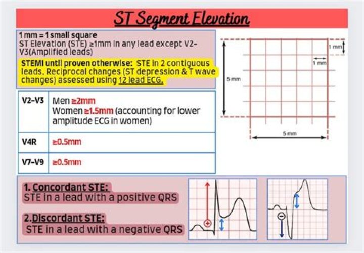
ST segment deviation (elevation, depression) is measured as the height difference (in millimeters) between the J point and the baseline (the PR segment). ST segment deviation occurs in a wide range of conditions, particularly acute myocardial ischemia.
What is normal ST segment elevation?
Thus, most men have elevation of the ST segment greater than 0.1 mV in the precordial leads. Therefore, elevation of the ST segment should be regarded as a normal finding and is often termed “male pattern”.
What ST elevation means?
ST-segment elevation myocardial infarction (STEMI) is the term cardiologists use to describe a classic heart attack. It is one type of myocardial infarction in which a part of the heart muscle (myocardium) has died due to the obstruction of blood supply to the area.
How do you find the ST segment?
The ST segment is the flat, isoelectric section of the ECG between the end of the S wave (the J point) and the beginning of the T wave. The ST Segment represents the interval between ventricular depolarization and repolarization.Where is J point measured?
The J point of the ECG is at the end of the QRS complex and the beginning of the ST segment. J point elevation can be seen in early repolarization.
What is V4 V5 V6 in ECG?
The electrical activity on an ECG (EKG). The areas represented on the ECG are summarized below: V1, V2 = RV. V3, V4 = septum. V5, V6 = L side of the heart.
What is a normal ST segment?
The ST segment is the interval between the end of the QRS complex (J point, or ST junction) and the beginning of the T wave. In the limb leads, the ST segment is isoelectric in about 75 percent of normal adults. 19. ST segment elevation or depression up to 0.1 mV generally is considered within normal limits.
Why is ST elevation in Michigan?
An acute ST-elevation myocardial infarction occurs due to occlusion of one or more coronary arteries, causing transmural myocardial ischemia which in turn results in myocardial injury or necrosis.How is ST elevation and depression measured?
ST segment deviation (elevation, depression) is measured as the height difference (in millimeters) between the J point and the baseline (the PR segment). ST segment deviation occurs in a wide range of conditions, particularly acute myocardial ischemia.
WHAT IS ST changes in ECG?ST and T wave changes may represent cardiac pathology or be a normal variant. Interpretation of the findings, therefore, depends on the clinical context and presence of similar findings on prior electrocardiograms. Nonspecific ST-T wave changes are very common and may be seen in any lead of the electrocardiogram.
Article first time published onHow do you read and interpret an ECG?
When interpreting the heart rhythm, you should look for P waves, which is a sign of atrial excitation. When every P wave is followed by a QRS complex, the ECG shows sinus rhythm. If the P waves are irregular, sinus arrhythmia is likely present.
How many mm of ST depression is significant?
ST segment depression may be determined by measuring the vertical distance between the patient’s trace and the isoelectric line at a location 2-3 millimeters from the QRS complex. It is significant if it is more than 1 mm in V5-V6, or 1.5 mm in AVF or III.
How is J point elevation different from ST elevation?
- There is elevation of the J point.
- The T wave is peaked and slightly asymmetrical.
- The ST segment and the ascending limb of the T wave form an upward concavity.
- The descending limb of the T wave is straighter and slightly steeper than the ascending limb.
What is ST elevation myocardial infarction?
ST-segment elevation myocardial infarction (STEMI) is the most acute manifestation of coronary artery disease and is associated with great morbidity and mortality. A complete thrombotic occlusion developing from an atherosclerotic plaque in an epicardial coronary vessel is the cause of STEMI in the majority of cases.
What is St level in TMT?
Criteria for Positive TMT Moderately positive : horizontal ST depression of 1.5 to 2.5 mm, slowly rising ST depression which remains depressed more than 2.5 mm 80 ms after the J point and down slopping ST depression with J point depressed 1.2 mm.
Where does V5 lead go?
V5 is placed directly between V4 and V6. V6 is placed over the fifth intercostal space at the mid-axillary line (as if drawing a line down from the armpit). V4-V6 should line up horizontally along the fifth intercostal space.
What does V1 V2 V3 mean?
At school, students often learn by heart the base, past simple and past participle (sometimes called V1, V2, V3, meaning Verb 1, Verb 2, Verb 3) for irregular verbs.
Are V1 V6 unipolar or bipolar?
The electrode leads each have a name. The bipolar extremity leads are called I, II and III. The unipolar extremity leads are called avR, avL and avF, and the chest leads are called V1–V6.
How do you analyze a ST segment?
ST segment depression is usually detected by choosing a reference point from the PR segment and a measurement point 60 or 80 ms after the J‐point. ST deviation is measured for each beat as the amplitude difference between these two points and mean values for 8 or 16 beats used for plotting and additional assessment.
Does STEMI cause VF?
The most common cause of pre-hospital death during STEMI is ventricular fibrillation; the widespread availability of automated external defibrillators, or AEDs, has been of benefit in this situation. Ventricular tachycardia also commonly occurs as well during and after STEMI and can be life-threatening.
Which is worse Nstemi or STEMI?
NSTEMI: What You Need to Know. NSTEMI stands for non-ST segment elevation myocardial infarction, which is a type of heart attack. Compared to the more common type of heart attack known as STEMI, an NSTEMI is typically less damaging to your heart.
Can a STEMI resolve itself?
Patients presenting with ST-elevation myocardial infarction (STEMI), whose symptoms and electrocardiographic changes completely resolve upon admission and before the administration of reperfusion therapy, pose a therapeutic dilemma.
When would ST elevation show on an ECG?
In an ECG recorded at a paper speed of 25 mm/s and an amplification of 10 mm/mV, the ST segment elevation from the baseline should be measured 80 ms after the J point and is considered present if the deviation is ≥0.2 mV in men and ≥0.15 mV in women in V2–V3 leads (≥0.1 mV in other leads).
Can you have ST elevation without MI?
To summarize, non-ischemic causes of ST-segment elevation include left ventricular hypertrophy, pericarditis, ventricular-paced rhythms, hypothermia, hyperkalemia and other electrolyte imbalances, and left ventricular aneurysm.
What are good ECG numbers?
The normal range of the ECG differed between men and women: heart rate 49 to 100 bpm vs. 55 to 108 bpm, P wave duration 81 to 130 ms vs. 84 to 130 ms, PR interval 119 to 210 ms vs.
What is a normal ECG reading?
Normal intervals Normal range 120 – 200 ms (3 – 5 small squares on ECG paper). QRS duration (measured from first deflection of QRS complex to end of QRS complex at isoelectric line). Normal range up to 120 ms (3 small squares on ECG paper).
What is P in ECG report?
The P wave and PR segment is an integral part of an electrocardiogram (ECG). It represents the electrical depolarization of the atria of the heart. It is typically a small positive deflection from the isoelectric baseline that occurs just before the QRS complex.
What are signs of ischemia on ECG?
The most common ECG sign of myocardial ischemia is flat or down-sloping ST-segment depression of 1.0 mm or greater. This report draws attention to other much less common, but possibly equally important, ECG manifestations of myocardial ischemia.
What is ST elevation in anterior leads?
The ECG findings of an acute anterior myocardial infarction wall include: ST segment elevation in the anterior leads (V3 and V4) at the J point and sometimes in the septal or lateral leads, depending on the extent of the MI. This ST segment elevation is concave downward and frequently overwhelms the T wave.
Is ST elevation bad?
Unlike skin or hair, once heart muscle is damaged, it will never grow back. All heart attacks are serious, but one type of is the most dangerous of all and it’s known as a STEMI (ST segment elevation myocardial infarction), or a widowmaker heart attack.
Can stress cause ST elevation?
They concluded that ST segment elevation is a good indicator of severe ischemia and poor collateral circulation. Several other case reports also found rare cases of ST elevations in non-Q wave leads during exercise stress testing that accurately predicted the presence of coronary stenoses (9–11).